
Cardiology Equipment
Advanced diagnosis and imaging for heart diseases is performed with ECG, exercise testing, echocardiography and bi-plane/mono-plane digital angiography systems.
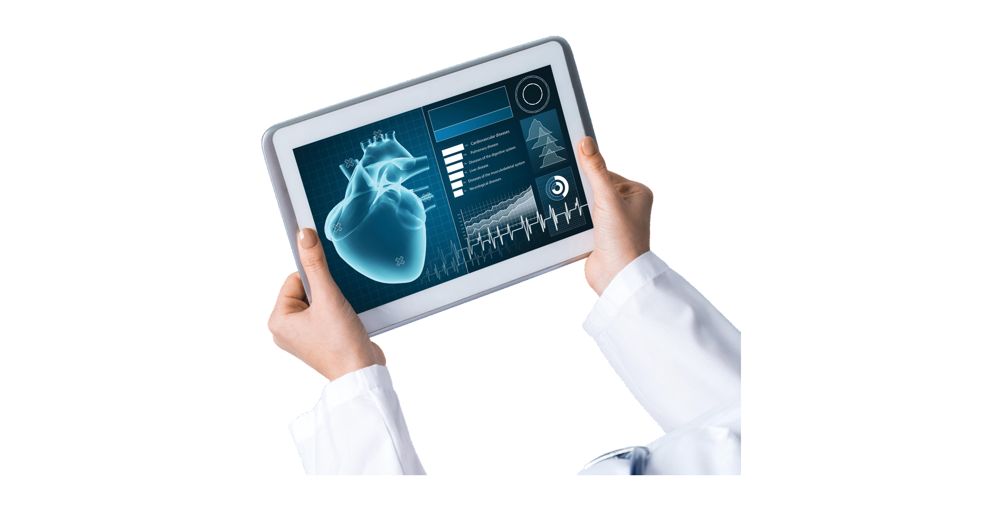
The Cardiology Department of Medipol Mega University Hospital provides services with world-class equipment and infrastructure to prevent, diagnose and treat heart diseases.
Examinations are performed 6 days a week (except Sunday) between 09:00 and 17:30 by specialists, associate professors, assistant professors and professors.
Planned patient journey
- 01 Online pre-consultation
- 02 Flight and welcome
- 03 Hospital appointment
- 04 Hotel and check-up
- 05 Return transfer
Treatment plan
Doctor's assessment and a category-specific procedure schedule.
Accommodation support
Discounted hotel and transfer times are planned together.
Get a second medical opinion
The opportunity to consult another specialist before deciding on treatment.
Multilingual communication
Receive service in your native language.

Contact Us
Leave a message.
When you fill out the form, your details are forwarded to the relevant email address.
The main methods used in the cardiology department for the prevention, diagnosis and treatment of heart and vascular diseases are summarised below. Click each heading to see a description of the relevant procedure.
Diagnostic Cardiology Examinations
Event Recorder
An event recorder is a device similar to an ECG Holter, used to capture infrequently recurring rhythm disorders. It can stay with the patient for a long period, and a recording taken at the moment of symptoms helps establish the diagnosis.
Electrocardiography (ECG)
An ECG is a basic examination that records the electrical activity of the heart to assess the conduction system of the heart muscle. It provides quick, painless information in the initial evaluation of rhythm disorders and many heart problems.
Blood Pressure Holter
A blood pressure Holter measures and records blood pressure and pulse at set intervals throughout the day. It is used to detect hidden hypertension and to monitor the effectiveness of treatment.
Exercise (Treadmill) Test
The exercise test evaluates how the heart responds to load at a gradually increasing pace on a treadmill. It is preferred for investigating coronary artery disease and measuring the effectiveness of treatment.
Transthoracic Echocardiography (ECHO)
Echocardiography is a method that examines the structure and function of the heart using sound waves (ultrasound). It involves no radiation, is painless, and provides extensive information for assessing the valves and chambers.
Three-Dimensional Transthoracic Echocardiography
Three-dimensional echocardiography images the heart valves and chambers in three dimensions. It offers clearer information, particularly for the detailed assessment of valve diseases.
Stress Echocardiography
Stress echocardiography examines changes in the contraction of the heart muscle while the workload of the heart is increased through exercise or medication. It is used to evaluate narrowing of the coronary arteries and valve problems.
Transoesophageal Echocardiography (TEE)
TEE is an advanced echocardiography method performed through the oesophagus when the chest structure does not provide adequate images. The valves, atria and any clots are imaged at higher resolution.
Three-Dimensional Transoesophageal Echocardiography (TEE)
Three-dimensional transoesophageal echocardiography provides three-dimensional detail for evaluating congenital heart defects, valve leaks and prosthetic valves. It is an advanced method performed in a limited number of centres in our country.
Rhythm (ECG) Holter
A rhythm Holter records the electrical activity of the heart, usually for 24 hours, via electrodes placed on the chest. It is effective at capturing rhythm disorders that vary throughout the day.
Tilt Table Test
The tilt test is used to diagnose fainting caused by a sudden drop in blood pressure or heart rate due to prolonged standing. The patient is monitored on a tilted table and the source of the complaints is investigated.
Interventional Procedures
Coronary Angiography
Coronary angiography is a diagnostic method that images narrowing or blockages in the vessels supplying the heart. Contrast agent is delivered into the vessels through a thin catheter and the coronary network is examined in detail.
Percutaneous Transluminal Coronary Angioplasty (PTCA) – Stent
Percutaneous coronary angioplasty is the procedure of opening a narrowing or blockage identified during angiography using a balloon and stent. The vessel is widened and blood flow restored without the need for open surgery.
Cardiac Catheterisation
Cardiac catheterisation, similar to coronary angiography, is performed through a vessel in the groin or arm, and intracardiac pressures are often measured as well. It provides information for the advanced assessment of valve and muscle diseases.
Opening Chronic Total Occlusions
In some patients who have had a heart attack, a vessel remains completely blocked. These vessels are opened using specialised interventional techniques requiring experience, restoring blood flow to the heart.
Non-Surgical Treatment of Aortic Stenosis (TAVI)
TAVI is the replacement of a narrowed aortic valve by catheter without open surgery. It offers a safe option especially for patients at high surgical risk.
Non-Surgical Treatment of Heart Defects
Holes in the heart that were once closed surgically can now, in suitable patients, be closed without surgery using occluder devices placed by catheter. Recovery time is shorter and the patient quickly returns to daily life.
Non-Surgical Vascular Repair
The endovascular method is the preferred option for patients at high risk for open surgery. It cannot be applied to every patient whose vascular anatomy is unsuitable, so the assessment is made by the physician.
A New Treatment for Resistant Hypertension: Renal Denervation
Around the renal arteries are sympathetic nerves that raise blood pressure. In renal denervation these nerves are deactivated by catheter, contributing to the control of resistant hypertension.
Nuclear Medicine Procedures
Myocardial Perfusion Scintigraphy (Thallium Test)
This test evaluates the blood supply of the heart muscle at rest and under load. It examines whether there is a blockage in the vessels supplying the heart.
Radiological Imaging
Coronary CT Angiography
Coronary CT angiography is an imaging method performed with a multi-slice tomography scanner, without a catheter and within a breath-hold. It is bloodless and painless, requires no hospital stay, and the patient returns to daily life after the procedure.
Cardiac MRI
Cardiac MRI is an imaging method that reveals diseases of the pericardium, heart chambers, valves, heart muscle and great vessels. The procedure takes about 45 minutes and requires no prior preparation.